Tracking phenotypes with automated lensfree microscopy
This project took place in winter term 2020, you CAN NOT apply to this project anymore!
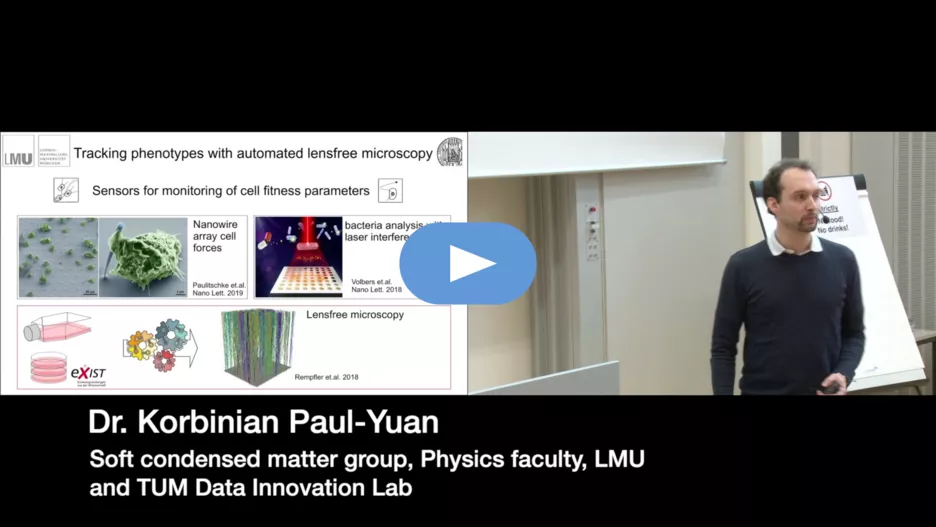
Results of this project are explained in the final presentation.
- Sponsored by: Soft condensed matter group, Physics faculty, LMU
- DI Incubator: Potential Startup
- Project Lead: Dr. Ricardo Acevedo Cabra
- Scientific Lead: Dr. Korbinian Paul-Yuan, Dr. Philipp Paulitschke
- TUM Co-Mentor: PhD candidate Michael Rauchensteiner

In vitro experiments with cultured cells are important in the first stage of developing more effective cancer treatments and to understand cellular mechanisms of cancer progression. By using microscopy and modern image detection algorithms the effect of potential drugs can be studied by quantifying growth and migration patterns. As part of our Exist-Transfer of Research project, financed by “Bundesministerium für Wirtschaft und Energie”, we have developed a super small, lensfree, digital holography microscope (LM) to extract phenotypic parameters in a non-invasive fashion. Moreover, the LM is much more scalable, can be universally integrated in cell biological experiments and will advance digitalization in the field of life science.
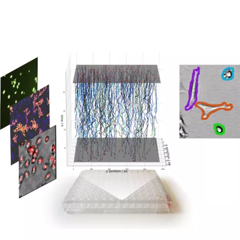
In publications [1,2] and a previous data-innovation-lab-project we have shown that our LM, equipped with smart image detection algorithms (deep convolutional neural networks, probabilistic models) can locate single cells, track their motion and segment clusters of cells (Figure) with the same quality as high resolution microscopy, which is ten times bigger and way more expensive.
The goal of this project is to assess the vital state of single cells noninvasively from lensfree microscopy images. Therefore, as an annotated dataset we prepared time laps videos using lensfree, bright field and fluorescence microscopy with various markers that label the vital state of a cell, for example to distinguish living from dead cells. The students can use the toolbox of previously developed image processing tools, extend it by tracking morphological changes of single cells to estimate the vital state of individual cells. This allows to analyze heterogeneities regarding drug response, detection of a phenotypical fingerprint of subpopulations and will allow more in-depth analysis of drug screening experiments. The result of this project will have direct impact in cell-biological labs, facilitating the work of cell biologists and will accelerate the foundation of the planned start-up.
References: [1] Rempfler M., S. Kumar, V. Stierle, P. Paulitschke, B. Andres and B.H. Menze. Cell Lineage Tracing in Lens-Free Microscopy Videos. Springer International Publishing, Cham; 2017:3–11. [2] Rempfler M., V. Stierle, K. Ditzel, S. Kumar, P. Paulitschke, B. Andres and B.H. Menze. (2018). Medical Image Analysis 2018; 48:147-161.

